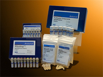
RiboAmp® Plus
 IComplete Solution for Gebe Expression Analysis
 |
1. Prepare Samples
|
|
 |
5. Microarray Labeling |
|

![]() Produce Micrograms from as Few as 250 Cells
Produce Micrograms from as Few as 250 Cells
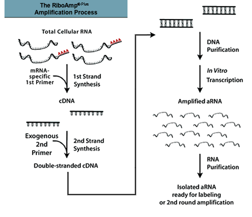
The RiboAmp Plus RNA Amplification Kit enables the researcher to perform molecular analysis on RNA samples that, until now, have been too small for microarray analysis. The RiboAmp Plus Kit offers unique linear amplification from as little as 1 ng of total RNA. Using a proprietary, patent- pending process for high-efficiency, high-fidelity linear amplification, messenger RNA is amplified up to 1000-fold in one synthesis round and up to 1,000,000-fold in two rounds. The RiboAmp Plus Kit produces amplified antisense RNA (aRNA), ready for labeling and microarray hybridization or QRT-PCR. The RiboAmp Plus Kit can be used to generate biologically relevant results with cDNA and oligonucleotide array platforms. The RiboAmp HS Plus kit is recommend for starting samples of 1ng of total RNA or less and 250 LCM cells or less.
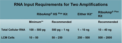
* In this range, the best kit to use depends upon the sample type and user preferences, since mRNA content varies among sample types.
** Minimum RNA input is the smallest amount tested in which useful levels of amplification can be achieved. Different sources of total RNA contain varying amounts of mRNA; consequently, the total RNA input needed to obtain microgram quantities of aRNA depends on the source of total RNA.
Maintain Native Expression Levels
The RiboAmp Plus RNA Amplification Kit offers unprecedented high-fidelity RNA amplification even from microscopic samples. Comparison of microarray results from total cellular RNA and from aRNA amplified using the RiboAmp Plus Kit reveal very high concordance between differentially expressed genes. This demonstrates the RiboAmp Plus Kit's ability to deliver the amplification fidelity needed for reliable data analysis from small samples.
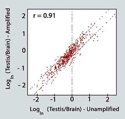
High correlation in differential expression between total cellular RNA and RiboAmp Plus amplified aRNA pairs. Total cellular RNA and aRNA samples amplified using the RiboAmp Plus Kit were generated from the same pool of mouse testis and brain samples, labeled and hybridized onto 9,000-element cDNA microarrays. Differential gene expression ratios between brain and testis were then calculated for the samples. The high correlation (r = 0.91) in differential gene expression between the amplified and unamplified samples indicates the high fidelity of the amplification process.
Detect Low-abundance mRNA
The RiboAmp Plus RNA Amplification Kit enables amplification of mRNA in all abundance classes. Using RT-PCR, low-, medium-, and high-abundance genes are readily detected within the amplified RNA population. Amplification of mRNA in all abundance classes ensures that differential gene expression patterns will be identified.
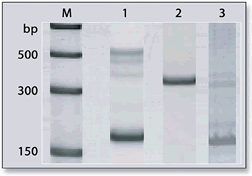
RT-PCR detection of low, medium, and high- abundance genes in amplified aRNA. Total cellular RNA was isolated from a mouse testis cell line, TM3, and amplified once using the RiboAmp Plus Kit. RT-PCR was then performed on three samples of the aRNA pool using three independent primer sets (Stratagene): Mouse Elongation Factor 1a (MEF-1a, high- abundance gene, ~3000 copies/cell, 187 bp fragment), Mouse Glyceraldehyde-3-Phosphate Dehydrogenase (M-GAPDH, medium- abundance gene, 300-3000 copies/cell, 357 bp fragment), and Mouse Protein Phosphatase 1 (MPP1, low-abundance gene, <300 copies/cell, 174 bp fragment). M: Marker. Lane 1: MEF-1a. Lane 2: M-GAPDH. Lane 3: MPP1.
Deliver Reproducible Amplified RNA for Microarray Experiments
The RiboAmp Plus RNA Amplification Kit consistently delivers aRNA ready for labeling and hybridization to expression microarrays with minimal amplification-to-amplification variability. Microarray analysis of independent amplifications from the same source RNA shows excellent correlation.
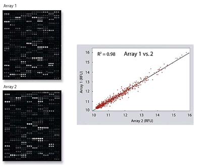
Microarray correlation of two independent amplifications. Total cellular RNA from a mouse testis cell line (TM3) was amplified using the RiboAmp Plus Kit. Three independent, one- round amplifications were performed from a single pool of RNA. Amplified aRNA was labeled with Cy5-dUTP and each labeled aRNA probe hybridized to a 9000 element mouse cDNA array (Arrays 1 and 2). Hybridized arrays were scanned and fluorescent intensities analyzed. One region of the two arrays is shown to demonstrate that similar gene expression patterns can be visualized. A scatter plot of relative fluorescent intensity (RFU) was generated to show correlation of independent amplification.
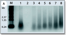
Amplification of aRNA from independent LCM samples. RNA isolated from six independent LCM captures from mouse kidney tissue and human breast cell line SKBR3 was amplified for two rounds with the RiboAmp Plus Kit, and run on an agarose gel. Typical aRNA lengths after two rounds of amplification range in size from below 250 to over 1,300 bases. M: Marker. Lane 1: positive amplification control (RiboAmp Plus Kit Control RNA). Lane 2, negative control. Lane 3: mouse kidney duct cells #1. Lane 4: mouse kidney duct cells #2. Lane 5: mouse kidney glomerulus. Lane 6: whole kidney. Lane 7: SKBR3 cell line (250 captured cells). Lane 8: SKBR3 cell line (500 captured cells).
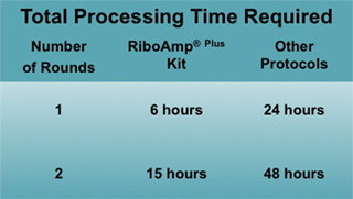
Process Samples Faster
The RiboAmp Plus Kit includes complete reagents and devices for template synthesis, transcription, and nucleic acid purification. Tedious vacuum concentrations are completely eliminated by incorporating optimized chemistries and by using Arcturus' novel, high-recovery MiraCol™ Purification Columns.
There are significantly fewer pipetting steps because the sample is eluted directly into reaction tubes and reagents are conveniently pre-dispensed and pre-mixed. In one eight-hour workday, the RiboAmp Plus Kit enables amplification, labeling, and microarray hybridization set up. With a convenient overnight in vitro transcription, a second round of amplification can be completed by the following morning.
![]() Reveal Differences in Gene Expression between Cell Types
Reveal Differences in Gene Expression between Cell Types
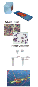
Accurate gene expression analysis requires the analysis of specific cell types without interference from surrounding cells. Starting with these pure cell populations often means working with small samples. Special technologies are needed to overcome the challenges of handling these precious samples. The combination of LCM, RNA amplification, and microarray analysis reveals differential gene expression between cell types.

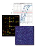
Panel A: Images showing normal ductal epithelium and invasive ductal carcinoma (IDC) from a human breast biopsy before and after Laser Capture Microdissection. Panel B: A GeneChip® Array (Affymetrix) image following hybridization of biotin-labeled aRNA generated from the normal and IDC breast samples. Panel C: Two-dimensional cluster analysis reveals reproducible patterns of up-regulation and repression of gene expression in cancer cells compared to normal, and more importantly, reveals stage-specific molecular signatures.
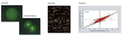
Total cellular RNA was isolated from two replicate samples of 20 mouse oocytes and 30 two-cell embryos using the PicoPure RNA Isolation Kit, amplified two rounds using the RiboAmp Plus RNA Amplification Kit, labeled and hybridized onto two separate 15,000-element cDNA microarrays to identify genes that are important in fertilization and pre-implantation development. Panel A: Mouse oocyte and embryo samples. Panel B: Micorarray hybridization image. Panel C: Scatter plot showing more than 450 genes differentially expressed between oocytes and embryos. Replicate hybridizations show very high correlation (R=0.89), suggesting excellent technical reproducibility of results obtained using amplified samples. Samples coutesy of A.J. Wyrobek, F.M. Marchetti and M. Coleman, Biology and Biotechnology Research Program, Lawrence Livermore National Laboratory, Livermore, CA, USA
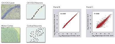
Panel A: Neurons from two distinct subtypes of a mouse brain, hippocampus CA1/CA2 and frontal cortex, were Laser Capture Microdissected using the PixCell IIe LCM System in replicate. The extracted RNA was purified with PicoPure RNA Isolation Kit, amplified using the RiboAmp Plus Kit, converted to labeled cDNA, and hybridized with two 23,040-element Mouse cDNA Microarrays (Agilent Technologies). Panel B: Microarray hybridizations of replicate samples of mouse hippocampus CA1/CA2 show very good correlation in Log2 relative fluorescent intensities (R=0.997), demonstrating high reproducibility of the entire process. Panel C: Comparison between mouse hippocampus CA1/CA2 and cortex shows differential expression of genes between the two neuron subtypes.
The RiboAmp RNA Plus Amplification Process
![]() Use Integrated Systems for Microgenomics
Use Integrated Systems for Microgenomics
Arcturus' complete System for Microgenomics offers an integrated solution for preparing small quantities of RNA for gene expression analysis. The System features Laser Capture Micro-dissection (LCM) with the ArcturusXT and Veritas Microdissection Instruments to procure pure cell populations, the PicoPure™ RNA Isolation Kit to extract and purify RNA, and the RiboAmp Plus Kit for linear amplification of RNA. Arcturus' Systems for Microgenomics enable accurate and sensitive microarray assays that reveal differential gene expression otherwise obscured in whole tissue samples or cell mixtures.
